ARDMS AE-Adult-Echocardiography Guaranteed Questions Answers - Practice AE-Adult-Echocardiography Test Online
Wiki Article
BONUS!!! Download part of TestkingPass AE-Adult-Echocardiography dumps for free: https://drive.google.com/open?id=1_rInwQSkm4MvzQq12sPQrnS1aLSzIMP2
More and more people look forward to getting the AE-Adult-Echocardiography certification by taking an exam. However, the exam is very difficult for a lot of people. Especially if you do not choose the correct study materials and find a suitable way, it will be more difficult for you to pass the exam and get the ARDMS related certification. If you want to get the related certification in an efficient method, please choose the AE-Adult-Echocardiography learning dumps from our company. We can guarantee that the study materials from our company will help you pass the exam and get the certification in a relaxed and efficient method.
ARDMS AE-Adult-Echocardiography Exam Syllabus Topics:
| Topic | Details |
|---|---|
| Topic 1 |
|
| Topic 2 |
|
| Topic 3 |
|
| Topic 4 |
|
| Topic 5 |
|
>> ARDMS AE-Adult-Echocardiography Guaranteed Questions Answers <<
Practice AE-Adult-Echocardiography Test Online & AE-Adult-Echocardiography Customized Lab Simulation
We know that tenet from the bottom of our heart, so all parts of service are made due to your interests. You are entitled to have full money back if you fail the exam even after getting our AE-Adult-Echocardiography test prep. Our staff will help you with genial attitude. We esteem your variant choices so all these versions of AE-Adult-Echocardiography Study Materials are made for your individual preference and inclination. Please get to know our AE-Adult-Echocardiography study materials as follows.
ARDMS AE Adult Echocardiography Examination Sample Questions (Q32-Q37):
NEW QUESTION # 32
Which finding is most consistent with this M-mode image?
- A. Rheumatic mitral stenosis
- B. Systolic antenor motion of the mitral valve
- C. Mitral valve annuloplasty ring
- D. Mitral valve prolapse
Answer: A
Explanation:
Comprehensive and Detailed Explanation From Exact Extract:
This M-mode echocardiographic image shows thickened mitral valve leaflets with a characteristic "doming" or "hockey-stick" appearance during diastole, which is classic for rheumatic mitral stenosis. Rheumatic mitral stenosis leads to leaflet thickening, restricted opening, and calcification, which alters the normal mitral valve motion on M-mode.
Mitral valve prolapse would show systolic displacement of the leaflets into the left atrium, typically later in systole, not doming in diastole. Mitral valve annuloplasty ring would appear as a bright echogenic line around the annulus but is not seen in this image. Systolic anterior motion (SAM) of the mitral valve is usually seen in hypertrophic cardiomyopathy and presents as anterior motion during systole, not the diastolic pattern shown.
This classical M-mode appearance is described in "Textbook of Clinical Echocardiography, 6e", Chapter on Rheumatic Valve Disease#20:385-390Textbook of Clinical Echocardiography#.
NEW QUESTION # 33
A continuous flow murmur is most likely due to which abnormality?
- A. Ebstein anomaly with atrial septal defect
- B. Ventricular septal defect
- C. Patent ductus arteriosus
- D. Concomitant aortic stenosis and mitral regurgitation
Answer: C
Explanation:
A continuous murmur, heard throughout systole and diastole, is most characteristically caused by a patent ductus arteriosus (PDA). PDA represents persistent communication between the aorta and pulmonary artery, allowing continuous blood flow during both phases of the cardiac cycle.
Ventricular septal defect usually produces a holosystolic murmur. Concomitant aortic stenosis and mitral regurgitation cause separate murmurs but not continuous. Ebstein anomaly with atrial septal defect typically produces murmurs related to tricuspid regurgitation or ASD but not a continuous murmur.
This clinical correlation is detailed in the "Textbook of Clinical Echocardiography, 6e", Chapter on Congenital Heart Disease and Murmur Etiologies#20:420-425Textbook of Clinical Echocardiography#.
NEW QUESTION # 34
Which finding is associated with partial anomalous venous return?
- A. Cleft mitral valve
- B. Persistent left superior vena cava
- C. Sinus venosus atrial septal defect
- D. Perimembranous ventricular septal defect
Answer: C
Explanation:
Partial anomalous pulmonary venous return (PAPVR) is a congenital defect where some pulmonary veins drain into the right atrium or systemic venous circulation rather than the left atrium. It is frequently associated with sinus venosus atrial septal defect (ASD), a defect near the junction of the superior vena cava and right atrium.
Cleft mitral valve is commonly associated with atrioventricular septal defects. Persistent left superior vena cava is a separate venous anomaly not typically linked with PAPVR. Perimembranous ventricular septal defects are different congenital defects not related to pulmonary venous anomalies.
The association between PAPVR and sinus venosus ASD is well described in the "Textbook of Clinical Echocardiography, 6e", Chapter on Congenital Heart Disease and Shunt Lesions#20:120-130Textbook of Clinical Echocardiography#
NEW QUESTION # 35
Which diagnosis is most likely confirmed by echocardiography in a 65-year-old female presenting with new onset chest pain associated with ST segment elevation on the electrocardiogram and angiographically normal coronary artenes?
- A. Apical hypertrophic cardiomyopathy
- B. Alcohol-associated cardiomyopathy
- C. Takotsubo cardiomyopathy
- D. Restrictive cardiomyopathy
Answer: C
Explanation:
Takotsubo cardiomyopathy, also known as stress-induced cardiomyopathy or "broken heart syndrome," predominantly affects postmenopausal women (usually older than 50 years) and often presents with acute chest pain and ST-segment elevation on the ECG mimicking acute myocardial infarction. However, coronary angiography reveals normal or non-obstructive coronary arteries.
Echocardiographically, Takotsubo cardiomyopathy is characterized by transient left ventricular systolic dysfunction with a typical pattern of apical ballooning and basal hyperkinesis. The wall motion abnormality extends beyond a single coronary artery territory, differentiating it from ischemic cardiomyopathy.
The diagnosis is supported by the clinical presentation, typical echocardiographic findings, and exclusion of obstructive coronary artery disease. The condition is usually reversible over days to weeks.
This is extensively described in the "Textbook of Clinical Echocardiography, 6e" (Chapter 8: Coronary Artery Disease and Takotsubo Syndrome), which highlights the typical patient demographics, presentation, echocardiographic features, and prognosis .
NEW QUESTION # 36
Which wall is indicated by the arrow on this image?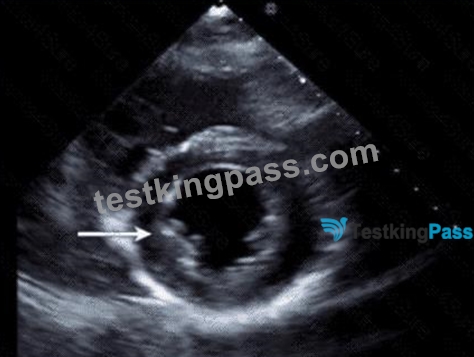
- A. Anterolateral
- B. Inferolateral
- C. Anterior
- D. Inferior
Answer: D
Explanation:
The echocardiographic image is a parasternal long axis or apical view showing the left ventricle. The arrow points to the wall segment located inferiorly, corresponding to the inferior wall of the left ventricle. The inferior wall is typically visualized in parasternal long axis and apical views as the posterior aspect of the ventricle.
Other options correspond to different walls: anterior is anterior septal wall, anterolateral and inferolateral refer to the lateral wall regions. Accurate wall identification is critical for regional wall motion analysis and coronary artery territory correlation.
This segmental wall identification is detailed in adult echocardiography and ASE chamber quantification guidelines#12:ASE Chamber Quantification Guidelinesp.90-95##16:Textbook of Clinical Echocardiography, 6ep.140-145#.
NEW QUESTION # 37
......
The TestkingPass AE Adult Echocardiography Examination (AE-Adult-Echocardiography) exam dumps are ready for quick download. Just choose the right AE-Adult-Echocardiography exam questions format and download it after paying an affordable AE Adult Echocardiography Examination in AE-Adult-Echocardiography Practice Questions charge and start this journey. Best of luck in the ARDMS AE-Adult-Echocardiography exam and career!!!
Practice AE-Adult-Echocardiography Test Online: https://www.testkingpass.com/AE-Adult-Echocardiography-testking-dumps.html
- Professional AE-Adult-Echocardiography Guaranteed Questions Answers - The Best Guide to help you pass AE-Adult-Echocardiography: AE Adult Echocardiography Examination ???? Open “ www.testkingpass.com ” and search for [ AE-Adult-Echocardiography ] to download exam materials for free ????New AE-Adult-Echocardiography Practice Questions
- AE-Adult-Echocardiography Study Guide ???? AE-Adult-Echocardiography Trustworthy Practice ???? Latest AE-Adult-Echocardiography Test Testking ???? Search for ➠ AE-Adult-Echocardiography ???? and obtain a free download on ▷ www.pdfvce.com ◁ ➰AE-Adult-Echocardiography Trustworthy Practice
- 2026 Useful AE-Adult-Echocardiography: AE Adult Echocardiography Examination Guaranteed Questions Answers ???? Easily obtain free download of ➤ AE-Adult-Echocardiography ⮘ by searching on [ www.testkingpass.com ] ????AE-Adult-Echocardiography Study Guide
- AE-Adult-Echocardiography Trustworthy Practice ???? AE-Adult-Echocardiography Reliable Study Questions ???? AE-Adult-Echocardiography Actual Exam Dumps ???? Open { www.pdfvce.com } and search for “ AE-Adult-Echocardiography ” to download exam materials for free ????AE-Adult-Echocardiography Reliable Study Questions
- Professional AE-Adult-Echocardiography Guaranteed Questions Answers - The Best Guide to help you pass AE-Adult-Echocardiography: AE Adult Echocardiography Examination ???? Open website 【 www.practicevce.com 】 and search for ➠ AE-Adult-Echocardiography ???? for free download ????AE-Adult-Echocardiography Reliable Exam Online
- Pass Guaranteed Quiz ARDMS - Accurate AE-Adult-Echocardiography Guaranteed Questions Answers ???? Go to website ▛ www.pdfvce.com ▟ open and search for ➡ AE-Adult-Echocardiography ️⬅️ to download for free ????AE-Adult-Echocardiography Positive Feedback
- www.verifieddumps.com Actual and Updated ARDMS AE-Adult-Echocardiography PDF Questions ???? [ www.verifieddumps.com ] is best website to obtain ☀ AE-Adult-Echocardiography ️☀️ for free download ????AE-Adult-Echocardiography Actual Exam Dumps
- AE-Adult-Echocardiography Valid Test Practice ???? AE-Adult-Echocardiography Reliable Dumps Book ???? AE-Adult-Echocardiography Reliable Study Questions ???? Search for ▛ AE-Adult-Echocardiography ▟ on 【 www.pdfvce.com 】 immediately to obtain a free download ????AE-Adult-Echocardiography Valid Exam Dumps
- High Hit-Rate AE-Adult-Echocardiography Guaranteed Questions Answers | AE-Adult-Echocardiography 100% Free Practice Test Online ???? Open ⏩ www.prep4away.com ⏪ enter ▷ AE-Adult-Echocardiography ◁ and obtain a free download ????AE-Adult-Echocardiography Exam Bible
- Newest AE-Adult-Echocardiography Guaranteed Questions Answers - Passing AE-Adult-Echocardiography Exam is No More a Challenging Task ???? ➥ www.pdfvce.com ???? is best website to obtain ▛ AE-Adult-Echocardiography ▟ for free download ????AE-Adult-Echocardiography Study Guide
- AE-Adult-Echocardiography Reliable Dumps Book ???? AE-Adult-Echocardiography Exam Bible ???? AE-Adult-Echocardiography Latest Exam Pattern ???? Download ➠ AE-Adult-Echocardiography ???? for free by simply entering { www.dumpsquestion.com } website ????Certification AE-Adult-Echocardiography Questions
- jeaneavp176574.wikibestproducts.com, tasneemchpu939339.blogdal.com, kalexiib247914.blog2freedom.com, luluaxrt715042.blogunteer.com, oisivxic615269.wikiparticularization.com, jessesvqs351538.blogpayz.com, camp-fire.jp, www.slideshare.net, fanniewckm111430.wikidirective.com, maenegh975523.nizarblog.com, Disposable vapes
P.S. Free 2026 ARDMS AE-Adult-Echocardiography dumps are available on Google Drive shared by TestkingPass: https://drive.google.com/open?id=1_rInwQSkm4MvzQq12sPQrnS1aLSzIMP2
Report this wiki page